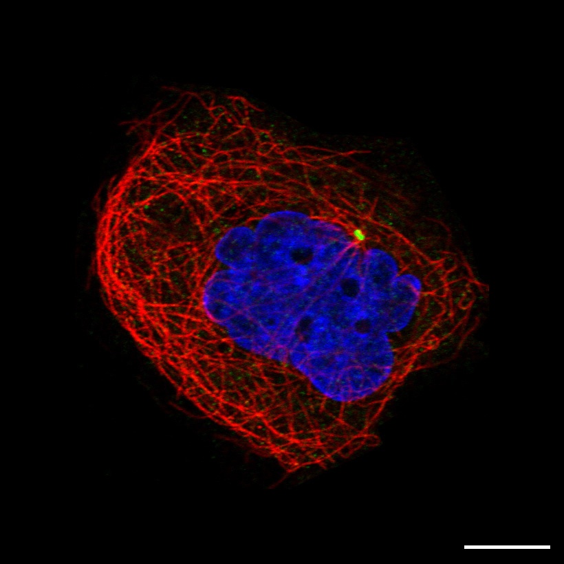
DictionaryCentrosomeCentrosomeScale bar represents 10µm CentrosomeThe centrosome is the main microtubule organizing center (MTOC) of the human cell. The centrosome consists of two centrioles that acts as scaffolds upon which the pericentriolar material is assembled. This fibrous material contains factors that nucleate and organize microtubules and are involved in eg. regulation cell cycle progression, mitosis, and cell polarity. Prior to cell division, the two centrioles move apart and replicates to form two complete centrosomes at opposite ends of the cell, where they direct formation of the bipolar mitotic spindle. Centriolar satellitesCentriolar satellites are small spherical granules with a diameter of 70-100 nm that cluster around the centrosome of human cells, as well as the basal body of ciliated cells. They consist of protein complexes of variable and dynamic composition, containing many proteins that also localize to centrosomes or basal bodies. Centriolar satellites are anchored to and move along microtubules with the help of motor proteins. Thus, they function as vehicles for protein transport towards, and likely also away from, the centrosome. Moreover, centriolar satellites seem to act as a hub for dynamic regulation of centrosome function and maintenance, as well as of ciliogenesis and neurogenesis, in response to a variety of signals. Indeed, both their content and dynamics seem to be subjected to regulatory control. Immunofluorescent stainingIt is difficult to clearly define the centrosome as it lacks a clear boundary towards the cytosol, and as it changes size as the amount of pericentriolar material varies during the cell cycle and between cell types. In the G1 phase, the centrosome lies at the center of the microtubules and is located close to the nuclear membrane. An antibody that specifically stains the two centrioles or a small single spot at the origin of microtubules is annotated as centrosome, whereas antibodies giving larger and more diffuse, sometimes punctate, staining of this area are annotated centriolar satellites. Also the size, abundance and location of centriolar satellites can var between cell types. Moreover, centriolar satellites undergo dissolution and are no longer visible upon entry into mitosis, but start to reappear after completion of cytokinesis. Read more about the proteome of the centrosome. |
The Project
The Human Protein Atlas
 The Human Protein Atlas project is funded


 MENU
MENU