Anatomy
Anatomy 1
Examples
Normal skeletal muscle
Protein expression
TNNT3, skeletal muscle DES, skeletal muscle DictionarySkeletal muscleSkeletal muscle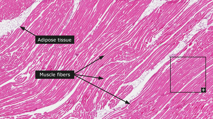
Skeletal muscleThe skeletal muscles and the heart muscle are composed of striated muscle tissue that form parallel muscle fibers. There are two types of muscle fibers (fast or slow) depending on the type of myosin present. These fiber types can not be distinguished in an ordinary HE staining. Development and normal activity of skeletal muscle are dependent and closely integrated with the nervous system. Skeletal muscles are attached to bone and contract voluntarily (via nerve stimulation) as opposed to the other common types of muscle, i.e. cardiac muscle and smooth muscle. The major cell type is myocytes that have been fused during development to form large multinucleated cells. The cells are rich in mitochondria and to a large extent contain actin and myosin proteins arranged in a repeating unit called a sarcomere. Histologically, this highly structured arrangement of sarcomeres appears as dark (A-bands) and light (I-bands) bands, which are clearly visible in the microscopic image. In addition to the muscle fibers, muscular tissues also consist of adjacent streaks of connective and adipose tissue. Muscle tissue is highly vascularized with a fine network of capillaries running in between the fibers. |
The Project
The Human Protein Atlas
 The Human Protein Atlas project is funded


 MENU
MENU


